ULTRASOUNDS
WHAT IS ULTRASOUND?
What is Ultrasound and how does it work?
Ultrasound or ultrasonography is a non-invasive diagnostic medical imaging technique that uses high frequency sound waves to produce images (sonogram) of the internal structures of the body. Frequencies above 20,000 Hz are called ultrasonic and the human ear cannot detect these sound waves. Ultrasound waves are produced by a transducer, which can both emit ultrasound waves, as well as detect the ultrasound echoes reflected. In most cases, the active elements in ultrasound transducers are made of special ceramic crystal materials called piezo- electric. This procedure is painless. Some discomfort may be felt in some instances, when the transducer is pressed against the skin for appropriate imaging. A standard scan takes approximately 30 – 60 minutes but please allow a little extra time as sometimes it can take a little longer than anticipated. Ultrasonography has a variety of uses in medical diagnostics. It is most well suited for imaging soft tissues that are solid and uniform or filled with fluid. In addition to other specialities, ultrasound is a very useful tool in detecting vascular problems with most of the smaller and larger blood vessels in the body. Using Doppler ultrasound technology, the flow of blood through the vessels can be observed and measured. It does not perform well when imaging calcified objects such as bone or objects filled with air like the bowel. Most ultrasonic exams are performed externally by running a transducer over the surface of the skin. Usually a gel is applied to the skin on which the transducer will glide during the exam. The gel helps prevent the formation of air pockets between the transducer and the skin that interfere with the ultrasonic signal. This ultrasound gel is water based, therefore when used as a coupling medium between transducer and body, helps in accurate assessment of depth and velocity measurements due to comparable sound velocities in soft tissue and water. Also help in quick drying from skin after cleaning the gel from skin.
Is ultrasound dangerous?
Ultrasound has been around for about 60 years now and studies have shown that it is a safe technique with no harmful side effects. Ultrasound imaging is not an X-ray as it uses sound waves and not ionizing radiation. Diagnostic ultrasound is a safe procedure that uses low-power sound waves. There are no known examples of tissue damage from conventional ultrasound imaging. Diagnostic ultrasound and/or sonography is considered a safe, non-invasive procedure by most every medical community. in part, because it uses low-power sound waves. No major medical source has proven any direct risks from a diagnostic ultrasound exam harmful enough to prevent its use. An ultrasound will not interact or cause any harm to a pacemaker.
Who will perform my scan?
Scans will be performed by an accredited medical sonographer who is fully qualified with suitable experience in performing the studies. A sonographer is a highly skilled medical imaging professional who uses ultrasound to perform specialised diagnostic examinations. The sonographer is legally required to scan the region requested on the referral from physician only. Ultrasound imaging is highly operator-dependent, and the outcome of a sonographic examination is dependent on the medical knowledge as well as the technical skills of the sonographer. During an ultrasound examination a sonographer will make real-time decisions to tailor the examination based on referral information, clinical context and the breadth of investigation required and selectively record anatomical images and physiological information that will form the basis of the clinical diagnosis A sonographer is trained in a post graduate level, specially to accurately perform an ultrasound examination and licensed for proper use of imaging equipment in a safe way. Sonographers understand ultrasound physics, cross sectional anatomy, physiology and pathology. Sonography requires specialized education and skills to view, analyse and modify the scan to optimize the information in the image. Because of the high levels of decisional latitude and diagnostic input, sonographers have a high degree of responsibility in the diagnostic process. They have core knowledge and skills in the applied anatomy, physiology and pathophysiology, application and operation of ultrasound imaging systems, ultrasound image recognition and comprehension, patient assessment, care and communication, critical thinking skills, ultrasound physics, occupational health and safety, infection control and quality assurance. Australian sonographers must be accredited by the Australian Sonographers Accreditation Registry (ASAR) and regularly monitored by continuing professional education.
Sonographer may ask to hold your breath – this is very important because when you breathe the organs go up and down in the tummy. When you hold your breath, the organs stay still allowing a better view of them.
WHAT WE OFFER

Abdominal Vascular Ultrasound
Sed ut perspiciatis unde omni na tus error sit volupt.

Carotid Arterial Duplex Ultrasound
Sed ut perspiciatis unde omni na tus error sit volupt.

Assessment for Deep and/or Superficial Thrombosis
Sed ut perspiciatis unde omni na tus error sit volupt.

Upper Limb Duplex Ultrasound
Sed ut perspiciatis unde omni na tus error sit volupt.

Lower Limb Arterial Duplex Ultrasound
Sed ut perspiciatis unde omni na tus error sit volupt.

Duplex Ultrasound scan to assess Varicose Veins
Sed ut perspiciatis unde omni na tus error sit volupt.

Upper Limb Duplex Ultrasound
An ultrasound scan may be necessary for your upper limbs. In this vascular laboratory we regularly assess for:
- venous and arterial assessment prior to arterio-venous fistulae for dialysis
- presence of deep or superficial thrombosis
- presence of arterial insufficiency
- review of arterio-venous fistulae for ongoing dialysis
This ultrasound is similar to the carotid arterial ultrasound. You will either be lying down or sitting up with your shirt removed enough to access the arm (from the shoulder down to the hand). You will be asked to remove any jewellery and/or watch. The sonographer will place gel on the examination region of the arm and place a probe (also known as a transducer) on the skin and image your vessels. The scan is non-invasive and painless. The test duration is 30 minutes.
Abdominal Vascular Ultrasound
An abdominal vascular ultrasound is similar to the leg arterial ultrasound to allow the sonographer to create a map detailing the location and extent of atheroma (plaque formation) or blockage (occlusion) or aneurysmal dilatation, by producing detailed images and blood flow data from the abdominal aorta and its major pelvic branches. In the case of veins, the map will detail the sites of leaking valves (incompetence) or clot.
This test is commonly performed for the assessment of:
- exercise-induced leg pain or suspicion of reduced pulses in the lower extremities.
- to assess the results of balloon angioplasty or stenting of the iliac arteries
- following EVAR (endovascular aneurysm repair), and sometimes open aneurysm repair.
- the arteries that branch off the abdominal aorta to supply the kidneys with blood.
- the arteries that branch off the abdominal aorta to supply blood to the stomach, intestines, colon, liver and spleen
- deep vein thrombosis (DVT).

In most cases of an abdominal vascular ultrasound requires you to fast for 6-8 hours . The examination is best performed in the early AM whilst fasting. Intestinal gas in the abdomen after eating is one of the major obstacles the sonographers face when performing this exam. We ask you to refrain from smoking and chewing gum or mints before the examination to facilitate better visualisation.
Diabetics should make the clerical staff aware of their condition at the time of booking, as staff will endeavour to make the appointment early in the morning, if this is possible.
All patients should take their usual oral medications with a small amount of water. Leave jewellery at home and wear loose, comfortable clothing. You may be asked to change into a gown.
The test takes about 45 – 60 minutes.

Assessment for Deep and/or Superficial Thrombosis
Venous ultrasound is used to detect blood clots), especially in the main veins of the leg – a condition often referred to as deep vein thrombosis. When duplex ultrasound is used to scan for a deep vein thrombosis (DVT) or thrombosis of the superficial veins (superficial thrombophlebitis) the ultrasound is used to check that the veins can be “squashed” by putting light pressure on them with the probe. Veins have thin walls so a normal vein can easily be squashed, and this appears as a “kissing” of the vein as it is pressed. However, a vein with thrombus within it is solid and cannot be compressed.
It is advisable to wear comfortable clothing for all venous ultrasound. You will be asked to remove some clothing to allow easy access to the area to be examined and you may be required to change into a gown. Assessing for deep venous thrombosis using duplex ultrasound is the same non-invasive test as for arterial and other venous ultrasound scans.
A water-soluble ultrasound gel is applied to the legs and a probe is run along the skin during the examination. The gel is easily washed off and does not stain clothes. The sonographer will scan you from the groin to the ankle (even though you have pain in the calf). The test duration is 15-30 minutes. If the sonographer detects a DVT, then you may require immediate treatment. The sonographer will notify the referring doctor with the results and you will be advised to see our specialist or your referring doctor after the test.
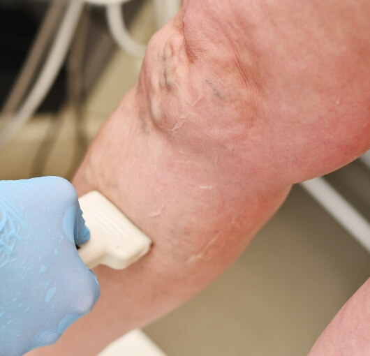
Duplex Ultrasound scan to assess Varicose Veins
A venous ultrasound study of the legs is performed to determine the cause of long-standing leg swelling, eczema, aching or pain or visible varicose veins. The term varicose veins is where the valves that normally keep blood flowing back to the heart may be damaged.
Varicose vein scans are usually performed standing up, with the weight on the leg that is not being scanned. In some cases, you may lie on a tilted couch instead of standing. In some cases, the leg may be examined whilst sitting on the edge of the couch with the leg dangling over the edge. The venous ultrasound is non-invasive and similar to the arterial ultrasounds with gel on the leg and running a probe along the skin. The sonographer will scan you from the groin to the ankle and the test duration is 30-60 minutes. The test should be painless. If you have any pain or discomfort or open wounds, please discuss this with the sonographer so they are aware for your comfort. To be able to assess the blood refluxing back down the veins, the sonographer may squeeze the calf or foot to stimulate the muscle pump. By examining the veins in the legs, a map of all the veins is produced for the surgeon to assess and plan the best course of treatment.

Lower Limb Arterial Duplex Ultrasound
An arterial duplex ultrasound is similar to the carotid ultrasound and is utilized to examine the circulation in the major blood vessels of the body. A map of the arteries in your abdomen and legs is detected by the sonographer to aid in the investigation of:
- Leg pain when walking
- Resting leg pain
- Foot, ankle, heel or toe ulcers
- Skin discolouration
- Aneurysm
- The results of balloon angioplasty, stenting or bypass operations.
The procedure is performed lying on the examination couch on your back.
The sonographer will apply warm gel to your abdomen and legs. You will be scanned from the level below your heart to your feet which will take about 30-60 minutes to complete.
A 6-8 hour fast may be required depending on the area to be assessed, clerical staff will advise you at the time of booking. Intestinal gas in the abdomen after eating is one of the major obstacles the sonographers face when performing this exam.
We ask you to refrain from smoking and chewing gum or mints before the examination to facilitate better visualisation.
Diabetics should make the clerical staff aware of their condition at the time of booking, as staff will endeavour to make the appointment early in the morning, if this is possible.
All patients should take their usual oral medications with a small amount of water.
Leave jewellery at home and wear loose, comfortable clothing. You may be asked to change into a gown.
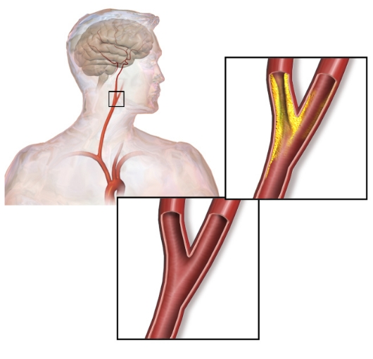
Carotid Arterial Duplex Ultrasound (or Carotid Doppler Ultrasound)
A carotid Doppler ultrasound is a non-invasive imaging test that uses ultrasound to examine the carotid arteries located in the neck. The duration of the test is 30 minutes. This test can show narrowing or possible blockages due to plaque build-up in the arteries which increases the risk of stroke. The results can help your doctor determine a treatment to lower your stroke risk. Wear a comfortable shirt with no collar or an open collar. Please don’t wear a necklace or dangling earrings in order to provide area in neck for imaging. A carotid ultrasound usually takes about 30 minutes. During the procedure, you’ll likely lie on your back during the ultrasound. The sonographer may position your head to better access the side of your neck with suitable pillow for extension. Warm gel will then be applied to your skin above the site of each carotid artery. The gel helps transmit the ultrasound waves back and forth. The sonographer then gently presses the transducer against the side of your neck. You shouldn’t feel any discomfort during the procedure. If you do, tell the sonographer. The sonographer will explain to you that you might hear some noise occasionally which is the Doppler ultrasound. Doppler is a physics term that allows the blood flow to be seen in colour and measured quantitatively. If your doctor ordered the carotid ultrasound as a follow-up to a surgical procedure, your doctor can explain whether the treatment is working, and whether you’ll need additional treatment or follow-up exams.